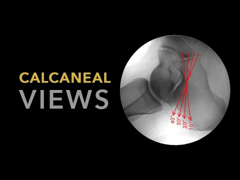
Plantar fasciitis is a common condition that causes heel pain and discomfort in the foot. But can you see it on an X-ray? Interestingly, plantar fasciitis does not typically show up on X-rays. This is because it is primarily an inflammation of the plantar fascia, the band of tissue that connects the heel bone to the toes. Unlike fractures or bone abnormalities, plantar fasciitis does not involve any structural changes in the bones themselves. Instead, it is a soft tissue injury that affects the ligaments and muscles in the foot.
While you may not be able to see plantar fasciitis on an X-ray, there are other ways to diagnose and treat this condition. Doctors often use a combination of physical exams, medical history, and symptoms to make a diagnosis. X-rays may still be useful in ruling out other conditions that can cause similar symptoms, such as stress fractures or bone spurs. Additionally, advanced imaging techniques like magnetic resonance imaging (MRI) or ultrasound can sometimes provide more detailed information about the soft tissues in the foot, allowing for a more accurate diagnosis.
Moving forward, let’s explore some key takeaways related to plantar fasciitis. We will discuss common risk factors, symptoms, and treatment options. Understanding these aspects can help individuals suffering from plantar fasciitis navigate their condition more effectively and seek the appropriate medical help. So, let’s dive into the world of plantar fasciitis and explore how to manage this debilitating foot condition.
key Takeaways
1. X-rays are not the most effective imaging method for diagnosing plantar fasciitis due to the fact that soft tissues like the plantar fascia cannot be clearly visualized.
2. X-rays can be useful in ruling out other potential causes of heel pain, such as stress fractures or bone spurs.
3. Diagnostic ultrasound is considered the most common imaging tool for diagnosing plantar fasciitis as it offers real-time visualization of soft tissue structures and can identify inflammation and thickening of the plantar fascia.
4. Magnetic resonance imaging (MRI) may be recommended if the diagnosis is still uncertain or for cases that do not respond to conservative treatment, as it can provide detailed information about soft tissue structures and identify associated conditions like nerve impingement or tumours.
5. A thorough physical examination, along with a detailed patient history, remains crucial in diagnosing plantar fasciitis, and imaging should be used as a supplementary tool to confirm the diagnosis and rule out other potential causes of heel pain.
Can Plantar Fasciitis Be Detected Using X-ray?
Understanding Plantar Fasciitis
Plantar fasciitis is a common foot condition characterized by inflammation and pain in the plantar fascia, a thick band of tissue that runs along the bottom of the foot. This condition often leads to heel pain, especially with the first steps after waking up or extended periods of rest. While clinical examination and patient history are often sufficient for diagnosing plantar fasciitis, some physicians may consider using additional imaging techniques such as X-rays to confirm the diagnosis.
The Role of X-ray Imaging
X-ray imaging plays a crucial role in determining the presence of underlying bone-related conditions and ruling out other potential causes of foot pain. Although plantar fasciitis primarily involves inflammation and damage to the soft tissues, X-rays can be helpful in identifying certain associated conditions, such as heel spurs, which are bony protrusions that can develop along the plantar fascia. However, it is important to note that the presence of a heel spur does not necessarily indicate the presence or severity of plantar fasciitis.
Limitations of X-ray for Diagnosing Plantar Fasciitis
While X-rays can provide valuable information, they are not the most effective tool for diagnosing plantar fasciitis itself. This is because X-ray images primarily focus on the bones and may not capture the inflammation and damage to the soft tissues that characterize plantar fasciitis. Therefore, a negative X-ray does not rule out the possibility of having plantar fasciitis, and a positive X-ray does not definitively confirm the diagnosis.
Alternative Imaging Techniques
When clinical symptoms and examination findings strongly suggest plantar fasciitis, X-rays are often unnecessary. However, in cases where the diagnosis is unclear or the healthcare professional needs to rule out other conditions, alternative imaging techniques may be considered. These may include ultrasound or magnetic resonance imaging (MRI) scans, which can provide more detailed visualization of the soft tissues and help identify abnormalities associated with plantar fasciitis.
Collaborative Approach to Diagnosis
The diagnosis of plantar fasciitis often requires a collaborative approach involving the patient, primary care physician, orthopedic specialist, and other healthcare professionals. While X-ray imaging can offer some insights, it should not be the sole basis for diagnosis. A comprehensive evaluation, including a thorough physical examination, reviewing medical history, and assessing the patient’s symptoms, is essential to accurately diagnose and treat plantar fasciitis.
Top 5 Tips for Diagnosing Plantar Fasciitis:
- Consult with a healthcare professional experienced in diagnosing foot conditions.
- Provide a detailed description of your symptoms, including the location and nature of the pain.
- Undergo a thorough physical examination to assess the signs of plantar fasciitis.
- Consider alternative imaging techniques like ultrasound or MRI if necessary.
- Follow the recommended treatment plan, which may include rest, stretching exercises, orthotics, and physical therapy.
Frequently Asked Questions
1. Can plantar fasciitis be detected through an X-ray?
Unfortunately, X-rays are not very effective in detecting plantar fasciitis. This is because plantar fasciitis is primarily a soft tissue condition, and X-rays are mainly used to visualize bones. However, X-rays can help rule out other potential causes of heel pain.
2. What diagnostic tools are used to confirm plantar fasciitis?
The diagnosis of plantar fasciitis is typically based on a physical examination, medical history, and the symptoms reported by the patient. In some cases, healthcare providers may request imaging tests like ultrasound or magnetic resonance imaging (MRI) to further evaluate the foot.
3. Are there any visible signs of plantar fasciitis on an X-ray?
No, there are no specific visible signs of plantar fasciitis on X-ray images. However, the X-ray may show other conditions such as heel spurs or stress fractures that could be contributing to the pain.
4. What can an X-ray show about heel spurs and plantar fasciitis?
An X-ray can sometimes reveal the presence of heel spurs, which are bony growths on the bottom of the heel bone. However, it’s important to note that heel spurs do not always cause symptoms or require treatment. Plantar fasciitis is usually caused by inflammation of the plantar fascia, the ligament that supports the arch of the foot.
5. Should I get an X-ray if I suspect plantar fasciitis?
An X-ray is not typically the first diagnostic test recommended for suspected plantar fasciitis. Healthcare providers generally start with a physical exam and obtain a thorough medical history to confirm the diagnosis. X-rays may be ordered if there is suspicion of other conditions or to rule out bone-related issues.
6. Can an X-ray help determine the severity of plantar fasciitis?
No, the severity of plantar fasciitis is not accurately determined through an X-ray. The diagnosis and severity assessment are primarily based on the patient’s reported symptoms, physical examination, and medical history.
7. Are there any risks associated with getting an X-ray for plantar fasciitis?
Generally, X-rays are safe and involve low levels of radiation. However, if you are pregnant or suspect you might be, it is essential to inform your healthcare provider. They will consider the potential risks and benefits before recommending any imaging procedures.
8. When are X-rays useful in diagnosing foot pain?
X-rays are useful when foot pain is suspected to be caused by conditions other than plantar fasciitis. They can help identify fractures, bone tumors, arthritis, and other bone-related issues that may contribute to the pain.
9. What are some alternative diagnostic methods for plantar fasciitis?
In addition to physical examinations, medical history, and imaging tests, healthcare providers may use provocative tests, such as the Windlass test or ultrasound elastography, to assess the condition of the plantar fascia and confirm the diagnosis of plantar fasciitis.
10. Can X-rays help monitor the progress of plantar fasciitis treatment?
No, X-rays are not typically used to monitor the progress of plantar fasciitis treatment. Improvement or resolution of symptoms is usually the main factor considered to evaluate treatment effectiveness.
Final Thoughts
When it comes to diagnosing plantar fasciitis, relying solely on X-rays may not provide conclusive results. Since plantar fasciitis is primarily a soft tissue condition, other diagnostic methods like physical examinations, medical history, and imaging tests such as ultrasounds are more frequently used. However, X-rays can still be useful in ruling out other potential causes of foot pain and identifying any accompanying bone-related issues.
If you’re experiencing symptoms of plantar fasciitis or persistent foot pain, it is recommended to consult a healthcare provider who can accurately assess your condition and recommend appropriate diagnostic tests and treatment options.

